3D Color X-Rays
3D Color X-Rays
By Sam Irani
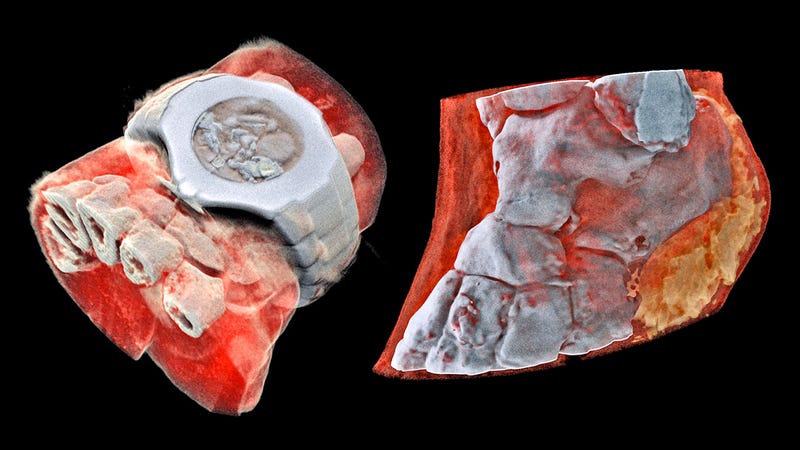
A couple months ago the first human was scanned with a revolutionary 3D color medical scanner developed by father and son scientists in New Zealand. Phil Butler, a physicist working at the University of Canterbury, and Anthony Butler, a radiologist at the Universities of Otago and Canterbury, invented the MARS spectral X-ray scanner, which has been commercialized by MARS Bioimaging.

The traditional approach to imaging the insides of a patient involves blasting them with x-rays. This electromagnetic radiation has a shorter wavelength than visible light, so it can easily pass through soft tissue, but it has more trouble passing through harder materials like bones. On the other side of your body, a sensor, or film, produces an image based on the intensity of the x-rays that make it through, thus revealing what’s inside you.
I think that the next steps from here are to find a way to spread the news due to the fact that this is so new. Another step might be to do more testing. From what I know there has not been that many people who have been tested. I think that in 4-5 years this technology will be the new standard for X-Rays and I think it could help a lot of people and make it easier for doctors to determine problems.
https://home.cern/news/news/knowledge-sharing/first-3d-colour-x-ray-human-using-cern-technology
https://www.engadget.com/2018/07/16/scientists-develop-world-first-3d-color-x-rays/
By Sam Irani
A couple months ago the first human was scanned with a revolutionary 3D color medical scanner developed by father and son scientists in New Zealand. Phil Butler, a physicist working at the University of Canterbury, and Anthony Butler, a radiologist at the Universities of Otago and Canterbury, invented the MARS spectral X-ray scanner, which has been commercialized by MARS Bioimaging.
The traditional approach to imaging the insides of a patient involves blasting them with x-rays. This electromagnetic radiation has a shorter wavelength than visible light, so it can easily pass through soft tissue, but it has more trouble passing through harder materials like bones. On the other side of your body, a sensor, or film, produces an image based on the intensity of the x-rays that make it through, thus revealing what’s inside you.
I think that the next steps from here are to find a way to spread the news due to the fact that this is so new. Another step might be to do more testing. From what I know there has not been that many people who have been tested. I think that in 4-5 years this technology will be the new standard for X-Rays and I think it could help a lot of people and make it easier for doctors to determine problems.
https://home.cern/news/news/knowledge-sharing/first-3d-colour-x-ray-human-using-cern-technology
https://www.engadget.com/2018/07/16/scientists-develop-world-first-3d-color-x-rays/


Comments
Post a Comment