Color X-Rays
Color X-Rays
By Luke Phillips
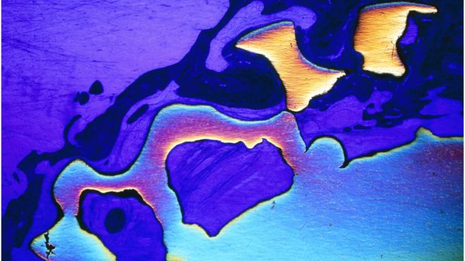
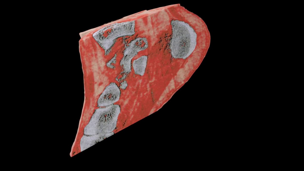
A couple months ago the first human was scanned with a revolutionary color medical X-ray scanner developed by a family of scientists in New Zealand. The scientists Phil Butler, a physicist working at the University of Canterbury, and Anthony Butler, a radiologist at the Universities of Otago and Canterbury, invented the MARS spectral X-ray scanner. This device was commercialized by MARS Bioimaging.

The traditional approach to imaging the insides of a patient involves blasting them with x-rays. This electromagnetic radiation has a shorter wavelength than visible light, so it can easily pass through soft tissue, but it has more trouble passing through harder organic materials like bones. On the other side of your body, a sensor produces an image based on the intensity of the x-rays that make it through, thus revealing what’s inside you. While this process is good at showing your bones it does not show a 3D view of them
I think that the next step is to make this device widespread. Another step might be to do more testing. From what I know there has not been that many people who have had the device used on them. In the not so distant future I believe this technology could revolutionize the medical field.
https://home.cern/news/news/knowledge-sharing/first-3d-colour-x-ray-human-using-cern-technology
https://www.engadget.com/2018/07/16/scientists-develop-world-first-3d-color-x-rays/